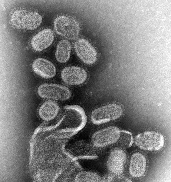
Archivo:EM of influenza virus.jpg
Apariencia

Tamaño de esta previsualización: 565 × 600 píxeles. Otras resoluciones: 226 × 240 píxeles · 452 × 480 píxeles · 700 × 743 píxeles.
Ver la imagen en su resolución original (700 × 743 píxeles; tamaño de archivo: 82 kB; tipo MIME: image/jpeg)
Historial del archivo
Haz clic sobre una fecha y hora para ver el archivo tal como apareció en ese momento.
| Fecha y hora | Miniatura | Dimensiones | Usuario | Comentario | |
|---|---|---|---|---|---|
| actual | 13:41 10 ago 2007 |  | 700 × 743 (82 kB) | ToNToNi | {{Information |Description=CDC, CDC Public Health Image Library (PHIL), http://phil.cdc.gov/Phil/details.asp |Source=Originally from [http://en.wikipedia.org en.wikipedia]; description page is/was [http://en.wikipedia.org/w/index.php?title=Image%3AEM_of_i |
Usos del archivo
No hay páginas que enlacen a este archivo.
Uso global del archivo
Las wikis siguientes utilizan este archivo:
- Uso en af.wikipedia.org
- Uso en an.wikipedia.org
- Uso en ar.wikipedia.org
- Uso en as.wikipedia.org
- Uso en awa.wikipedia.org
- Uso en azb.wikipedia.org
- Uso en az.wikipedia.org
- Uso en bat-smg.wikipedia.org
- Uso en ba.wikipedia.org
- Uso en be-tarask.wikipedia.org
- Uso en be.wikipedia.org
- Uso en bg.wikipedia.org
- Uso en bn.wikipedia.org
- Uso en bo.wikipedia.org
- Uso en br.wikipedia.org
- Uso en bs.wikipedia.org
- Uso en bxr.wikipedia.org
- Uso en ca.wikipedia.org
- Uso en cdo.wikipedia.org
- Uso en ckb.wikipedia.org
- Uso en csb.wikipedia.org
- Uso en cs.wikipedia.org
- Uso en da.wikipedia.org
- Uso en de.wikipedia.org
- Uso en en.wikipedia.org
- Avian influenza
- Emergent virus
- Portal:Medicine/Selected Article Archive
- Wikipedia:Today's featured article/January 2007
- Wikipedia:Today's featured article/January 1, 2007
- Portal:Medicine/Selected article/8, 2008
- Portal:Medicine/Selected Article
- Portal:Medicine/Selected Article/10
- Influenza
- Wikipedia:VideoWiki/Influenza
- User:JenOttawa/Notes/practice
- Uso en en.wikibooks.org
- Uso en en.wikinews.org
- Uso en et.wikipedia.org
- Uso en eu.wikipedia.org
- Uso en fa.wikipedia.org
Ver más uso global de este archivo.


