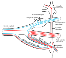
Célula de Dogiel
Apariencia

Las células de Dogiel, son un tipo de neuronas multipolares[1] que se encuentran dentro de los ganglios simpáticos prevertebrales.[2][3][4] Se conocen así por el anatomista y fisiólogo ruso Alexandre Dogiel (1852–1922).[5] Desempeñan un papel en el sistema nervioso entérico.[6][7]
Tipos
[editar]Existen siete tipos de células de Dogiel.[6][8]
Referencias
[editar]- ↑ Brookes, SJ; Song, ZM; Ramsay, GA; Costa, M (May 1995). «Long aboral projections of Dogiel type II, AH neurons within the myenteric plexus of the guinea pig small intestine.». The Journal of Neuroscience 15 (5 Pt 2): 4013-22. PMID 7751962.
- ↑ Cajal, Santiago R.y; Pasik, Pedro; Pasik, Tauba (14 de octubre de 2002). Texture of the Nervous System of Man and the Vertebrates. Springer Science & Business Media. p. 5. ISBN 978-3-211-83202-8.
- ↑ «Gray, Henry. 1918. Anatomy of the Human Body. Page 921». www.bartleby.com. Consultado el 5 de noviembre de 2020.
- ↑ Barrett, Kim E.; Ghishan, Fayez K.; Merchant, Juanita L.; Hamid M. Said; Jackie D. Wood (10 de mayo de 2006). Physiology of the Gastrointestinal Tract. Academic Press. p. 617. ISBN 978-0-08-045615-7.
- ↑ «Dogiel cells». Medical Eponyms. Medical Eponyms.
- ↑ a b Furness, John Barton (15 de abril de 2008). The Enteric Nervous System. John Wiley & Sons. pp. 35-38. ISBN 978-1-4051-7344-5.
- ↑ Stach, W (1979). «[Differentiated vascularization of Dogiel's cell types and the preferred vascularization of type I/2 cells within plexus myentericus (Auerbach) ganglia of the pig (author's transl)].». Anatomischer Anzeiger (en alemán) 145 (5): 464-73. PMID 507375.
- ↑ Scheuermann, DW; Stach, W (1983). «External adrenergic innervation of the three neuron types of Dogiel in the plexus myentericus and the plexus submucosus externus of the porcine small intestine.». Histochemistry 77 (3): 303-11. PMID 6863029. doi:10.1007/BF00490893.
- Lectura complementaria
- Furness, John Barton (15 de abril de 2008). The Enteric Nervous System. John Wiley & Sons. pp. 35-38. ISBN 978-1-4051-7344-5.
- Tache, Yvette; Wingate, David L.; Burks, Thomas F. (2 de diciembre de 1993). Innervation of the Gut. CRC Press. p. 268. ISBN 978-0-8493-4718-4.
- Stach, W (1979). «[Differentiated vascularization of Dogiel's cell types and the preferred vascularization of type I/2 cells within plexus myentericus (Auerbach) ganglia of the pig (author's transl)].». Anatomischer Anzeiger (en alemán) 145 (5): 464-73. PMID 507375.
- Cornelissen, WB; de Laet, AB; Kroese, AB; Adriaensen, DW; van Bogaert, PP; Scheuermann, DW; Timmermans, JP (Oct 1999). «Species-dependent features of Dogiel type II neurones in the mammalian enteric nervous system.». European Journal of Morphology 37 (4–5): 241-9. PMID 10477469. doi:10.1076/ejom.37.4.241.4730.
