
Archivo:Anaplastic astrocytoma.jpg
Tamaño de esta previsualización: 800 × 552 píxeles. Otras resoluciones: 320 × 221 píxeles · 640 × 442 píxeles · 1024 × 707 píxeles · 1200 × 828 píxeles.
Ver la imagen en su resolución original (1200 × 828 píxeles; tamaño de archivo: 200 kB; tipo MIME: image/jpeg)
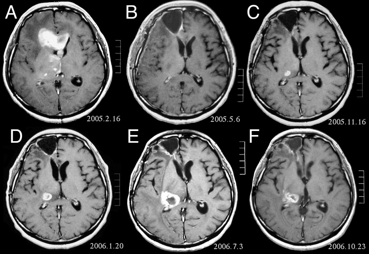
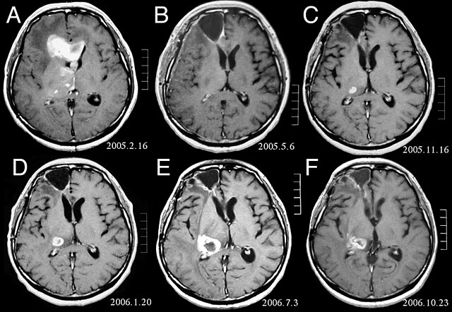
| Descripción | MRI of brain. (A) Initial MRI on February 16, 2005, shows a tumor in the right and left frontal lobe as well as the right thalamus. (B) MRI after surgery, radiation and chemotherapy. The tumor has completely disappeared except for slight enhancement adjacent to the surgical margin. (C) Recurrence of the thalamic tumor despite maintenance chemotherapy on November 16, 2005. (D) Increase in size of the thalamic tumor two months after stereotactic radiotherapy. (E) After 6 cycles of TMZ therapy, the thalamic lesion enlarged, and the patient developed dysarthria and hemiparesis. (F) After 2 courses of treatment with interferon-beta and TMZ, the tumor shows a partial response. |
| Fecha | |
| Fuente | Fujimaki T, Ishii H, Matsuno A, Arai H, Nakagomi T.Effectiveness of interferon-beta and temozolomide combination therapy against temozolomide-refractory recurrent anaplastic astrocytoma.World J Surg Oncol. 2007 Aug 4;5:89. PMID 17683572 doi:10.1186/1477-7819-5-89 |
| Autor | Fujimaki T, Ishii H, Matsuno A, Arai H, Nakagomi T. |
| Permiso (Reutilización de este archivo) |
BioMedCentral License |
Este archivo está disponible bajo la licencia Creative Commons Atribución 2.0 Genérica.
- Eres libre:
- de compartir – de copiar, distribuir y transmitir el trabajo
- de remezclar – de adaptar el trabajo
- Bajo las siguientes condiciones:
- atribución – Debes otorgar el crédito correspondiente, proporcionar un enlace a la licencia e indicar si realizaste algún cambio. Puedes hacerlo de cualquier manera razonable pero no de manera que sugiera que el licenciante te respalda a ti o al uso que hagas del trabajo.
Historial del archivo
Haz clic sobre una fecha y hora para ver el archivo tal como apareció en ese momento.
| Fecha y hora | Miniatura | Dimensiones | Usuario | Comentario | |
|---|---|---|---|---|---|
| actual | 16:47 25 feb 2008 | | 1200 × 828 (200 kB) | Filip em | {{Information |Description=MRI of brain. (A) Initial MRI on February 16, 2005, shows a tumor in the right and left frontal lobe as well as the right thalamus. (B) MRI after surgery, radiation and chemotherapy. The tumor has completely disappeared except f |
Usos del archivo
La siguiente página usa este archivo:
Uso global del archivo
Las wikis siguientes utilizan este archivo:
- Uso en ar.wikipedia.org
- Uso en bg.wikipedia.org
- Uso en cs.wikipedia.org
- Uso en da.wikipedia.org
- Uso en de.wikipedia.org
- Uso en el.wikipedia.org
- Uso en en.wikipedia.org
- Uso en et.wikipedia.org
- Uso en hi.wikipedia.org
- Uso en hr.wikipedia.org
- Uso en hu.wikipedia.org
- Uso en it.wikipedia.org
- Uso en kk.wikipedia.org
- Uso en lb.wikipedia.org
- Uso en lv.wikipedia.org
- Uso en mk.wikipedia.org
- Uso en mt.wikipedia.org
- Uso en nl.wikipedia.org
- Uso en no.wikipedia.org
- Uso en pl.wikipedia.org
- Uso en ro.wikipedia.org
- Uso en sk.wikipedia.org
- Uso en sl.wikipedia.org
- Uso en sq.wikipedia.org
- Uso en sr.wikipedia.org
- Uso en uk.wikipedia.org