Diferencia entre revisiones de «Vía rostral migratoria»
Sin resumen de edición |
Sin resumen de edición |
||
| Línea 1: | Línea 1: | ||
{{Infobox |
{{Infobox Brain |
||
| Name |
| Name = Subventricular Zone |
||
| NeuroLex = Subventricular Zone |
|||
| Latin = |
|||
| NeuroLexID = nlx_144262 |
|||
| GraySubject = 187 |
|||
| GrayPage = 791 |
|||
| Image = CerebellumDiv.png |
|||
| Caption = Schematic representation of the major anatomical subdivisions of the cerebellum. Superior view of an "unrolled" cerebellum, placing the vermis in one plane. |
|||
| Image2 = Gray703.png |
|||
| Caption2 = Anterior view of the [[cerebellum]]. ("Flocculus" labeled at upper right.) |
|||
| IsPartOf = [[Cerebellum]] |
|||
| Components = |
|||
| Artery = [[Anterior inferior cerebellar artery|AICA]] |
|||
| Vein = |
|||
| System = [[Vestibular system|Vestibular]] |
|||
| BrainInfoType = hier |
|||
| BrainInfoNumber = 677 |
|||
| MeshName = |
|||
| MeshNumber = |
|||
| NeuroLex = Flocculus |
|||
| NeuroLexID = birnlex_1329 |
|||
| DorlandsPre = f_09 |
|||
| DorlandsSuf = 12368554 |
|||
}} |
}} |
||
The '''flocculus''' ([[Latin]]: ''tuft of wool'', diminutive) is a small lobe of the [[cerebellum]] at the posterior border of the middle cerebellar peduncle anterior to the [[biventer lobule]]. Like other parts of the cerebellum, the flocculus is involved in motor control. It is an essential part of the [[vestibulo-ocular reflex]], and aids in the learning of basic motor skills in the brain. |
|||
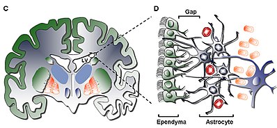
[[Archivo:Human_subventricular_zone.jpg|miniaturadeimagen|400x400px|Zona subventricular humana. Imagen de Oscar Arias-Carrión, 2008.]] |
|||
It is associated with the nodulus of the [[vermis]]; together, these two structures compose the vestibular part of the cerebellum. |
|||
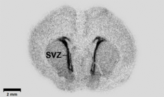
[[Archivo:Autoradiography_of_a_brain_slice_from_an_embryonal_rat_-_PMID19190758_PLoS_0004371.png|miniaturadeimagen|240x240px|En un cerebro de rata en estadio embrionario, GAD67, marcador qeue tiende a concentrarse en la SVZ. imágen de Popp et al., 2009.<ref name="pmid19190758">{{cite journal |vauthors=Popp A, Urbach A, Witte OW, Frahm C |title=Adult and Embryonic GAD Transcripts Are Spatiotemporally Regulated during Postnatal Development in the Rat Brain |journal=[[PLoS ONE]] |volume=4 |issue=2 |pages=e4371 |year=2009 |pmid=19190758 |pmc=2629816 |doi=10.1371/journal.pone.0004371 |url=http://dx.plos.org/10.1371/journal.pone.0004371 |editor1-last=Reh |editor1-first=Thomas A.}}</ref>]] |
|||
La '''zona subventricular''' ('''SVZ''') es un término utilizado para describir tanto el tejido embrionario como el tejido neuronal adulto del [[sistema nervioso central]] de los [[Vertebrata|vertebrados]] (SNC) localizado bajo los [[Ventrículos laterales|Ventrículos laterales.]] En la etapa embrionaria, la SVZ refiere a una zona secundaria de proliferación que contiene células progenitoras neurales que se dividen para producir neuronas en el proceso llamado [[neurogénesis]].<ref>{{cite journal|last1=Noctor|first1=SC|last2=Martínez-Cerdeño|first2=V|last3=Ivic|first3=L|last4=Kriegstein|first4=AR|title=Cortical neurons arise in symmetric and asymmetric division zones and migrate through specific phases.|journal=Nature Neuroscience|date=February 2004|volume=7|issue=2|pages=136–44|pmid=14703572|doi=10.1038/nn1172}}</ref> Las células madre neurales primarias del [[Encéfalo]] y la [[Médula espinal]], llamadas células gliales radiales, residen en la zona ventricular (VZ) (llamada así porque la VZ delimita los ventrículos en el desarrollo).<ref name="ReferenceA">{{cite journal|last1=Rakic|first1=P|title=Evolution of the neocortex: a perspective from developmental biology.|journal=Nature reviews. Neuroscience|date=October 2009|volume=10|issue=10|pages=724–35|pmid=19763105|doi=10.1038/nrn2719|pmc=2913577}}</ref> En la corteza cerebral en desarrollo del telencéfalo dorsal la SVZ y la VZ son tejidos transitorios que no existen en el adulto.<ref name="ReferenceA"/> En cambio, la SVZ del telencéfalo ventral persiste a lo largo de la vida. |
|||
At its base, the flocculus receives input from the inner ear's vestibular system and regulates balance. Many floccular projections connect to the motor nuclei involved in control of eye movement. |
|||
La SVZ adulta es una estructura apareada del encéfalo que se sitúa a a lo largo de las paredes laterales de los ventrículos laterales.<ref name="Quinones-Hinojosa, A. 2006">{{cite journal|last=Quiñones-Hinojosa|first=A|author2=Sanai, N |author3=Soriano-Navarro, M |author4=Gonzalez-Perez, O |author5=Mirzadeh, Z |author6=Gil-Perotin, S |author7=Romero-Rodriguez, R |author8=Berger, MS |author9=Garcia-Verdugo, JM |author10= Alvarez-Buylla, A |title=Cellular composition and cytoarchitecture of the adult human subventricular zone: a niche of neural stem cells.|journal=The Journal of Comparative Neurology|date=Jan 20, 2006|volume=494|issue=3|pages=415–34|doi=10.1002/cne.20798|pmid=16320258}}</ref> Está compuesta de cuatro capas distintas de grosor, densidad y composición celular celular variable.<ref name="multiple">{{cite journal|last=Quiñones-Hinojosa|first=A|author2=Chaichana, K |title=The human subventricular zone: a source of new cells and a potential source of brain tumors.|journal=Experimental neurology|date=Jun 2007|volume=205|issue=2|pages=313–24|doi=10.1016/j.expneurol.2007.03.016|pmid=17459377}}</ref> Junto con el [[giro dentado]] del [[Hipocampo (anatomía)|hipocampo]] la SVZ es uno de los dos sitios donde se ha encontrado [[neurogénesis]] en el cerebro de mamíferos adultos.<ref name="Ming 2011">{{cite journal|last=Ming|first=GL|author2=Song, H |title=Adult neurogenesis in the mammalian brain: significant answers and significant questions.|journal=Neuron|date=May 26, 2011|volume=70|issue=4|pages=687–702|doi=10.1016/j.neuron.2011.05.001|pmid=21609825|pmc=3106107}}</ref> |
|||
== Structure== |
|||
The flocculus is contained within the flocculonodular lobe which is connected to the cerebellum. The cerebellum is the section of the brain that is essential for [[motor control]]. As a part of the cerebellum, the flocculus plays a part of the vestibulo-ocular reflex system, a system that controls the movement of the eye in coordination with movements of the head.<ref name=Purves/> There are five separate “zones” in the flocculus and two halves, the caudal and rostral half. |
|||
== Estructura == |
|||
===Circuitry of the flocculus=== |
|||
The flocculus has a complex circuitry that is reflected in the structure of the zones and halves. These "zones" of the flocculus refer to five separate groupings of [[Purkinje cells]] that project to different areas of the brain. Depending upon where stimulus occurs in the flocculus, signals can be projected to very different parts of the brain. The first and third zones of the flocculus project to the superior vestibular nucleus, the second and fourth zone projects to the medial vestibular nucleus, and the fifth zone projects to the interposed posterior nucleus, a part of the cerebellum.<ref>{{cite journal |doi=10.1002/cne.903490308 |pmid=7852634 |title=Projections of individual purkinje cells of identified zones in the flocculus to the vestibular and cerebellar nuclei in the rabbit |journal=The Journal of Comparative Neurology |volume=349 |issue=3 |pages=428–47 |year=1994 |last1=De Zeeuw |first1=C. I. |last2=Wylie |first2=D. R. |last3=Digiorgi |first3=P. L. |last4=Simpson |first4=J. I. }}</ref> |
|||
=== Capa I === |
|||
The anatomy of the flocculus shows that it is composed of two disjointed lobes or halves. The “halves” of the flocculus refer to the caudal half and the rostral half, and they indicate from where fiber projections are received and the path in which a signal travels.<ref name=Ito>{{cite journal |doi=10.1146/annurev.ne.05.030182.001423 |pmid=6803651 |title=Cerebellar Control of the Vestibulo-Ocular Reflex--Around the Flocculus Hypothesis |journal=Annual Review of Neuroscience |volume=5 |pages=275–96 |year=1982 |last1=Ito |first1=M }}</ref> The caudal half of the flocculus receives [[mossy fiber (cerebellum)|mossy fiber]] projections mainly from the [[vestibular system]] and nucleus reticularis tegmenti pontis, an area within the floor of the midbrain that affects the axonal projections or images received by the cerebellum. Vestibular inputs are also carried through [[climbing fibers]] that project into the flocculus, stimulating Purkinje cells. Leading research would suggest that climbing fibers play a specific role in motor learning.<ref name=Lisberger>{{cite journal |doi=10.1126/science.3055293 |pmid=3055293 |title=The neural basis for learning of simple motor skills |journal=Science |volume=242 |issue=4879 |pages=728–35 |year=1988 |last1=Lisberger |first1=S. |bibcode=1988Sci...242..728L }}</ref> The climbing fibers then send the image or projection to the part of the brain that receives electrical signals and generates movement. From the midbrain, [[Corticopontine fibers]] carry information from the primary motor cortex.<ref name=Purves/> From there, projections are sent to the [[ipsilateral]] [[pontine nucleus]] in the ventral pons, both of which are associated with projections to the cerebellum. Finally, pontocerebellar projections carry vestibulo-occular signals to the contralateral cerebellum via the middle cerebellar peduncle.<ref>{{cite web |last=McDougal |first= David |last2=Van Lieshout |first2= Dave |last3=Harting |first3=John |title= Pontine Nuclei and Middle Cerebellar Penduncle |url=http://www.neuroanatomy.wisc.edu/virtualbrain/BrainStem/16Pontine.html |accessdate = 28 April 2013}}</ref> The rostral half of the flocculus also receives mossy fiber projections from the [[pontine nuclei]]; however, it receives very little projection from the vestibular system. |
|||
La capa interior (Capa I) contiene una monocapa de células ependimales que revisten la cavidad ventricular; estas células tienen un solo cíluo apical y muchas expansiones basales que pueden estar tanto paralelas como perpendiculares a la superficie ventricular. Estas expansiones pueden interaccionar intimamente con los procesos astrocíticos que las conectan con la capa hipocelular (Capa II).<ref name="multiple" /> |
|||
== |
=== Capa II === |
||
La capa secundaria (Capa II) proporciona for a hypocellular gap abutting the former" y ha sido demostrado que contiene una red funcional de procesos astrocíticos relacionados entre sí y que expresan GFAP. Estos procesos forman complejos funcionales a pesar de que esta zona carece de cuerpo celulares a excepción de algún soma neuronal aislado. La función de esta capa es desconocida en humanos sin embargo se ha hipotetizado que las conexiones entre astrocitos y las células ependimales entre las capas I y II podrían estar regulando funciones neuronales como la homeostasis metabólica y/o el control de la proliferación de las células madre así como su diferenciación durante el desarrollo. Potencialmente, tales características podrían hacer que actuase como un resto del desarrollo temprano o una vía para la migración celular dada su similitud con una capa homóloga en la SVZ bovina que también tiene células migratorias, comunes solo en ordenes de animales superiores.<ref name="multiple" /> |
|||
The flocculus is a part of the vestibulo-ocular reflex system and is used to help stabilize gaze during head rotation about any axis of space. Neurons in both the vermis of cerebellum and flocculus transmit an eye velocity signal that correlates with [[smooth pursuit]]. |
|||
=== Capa III === |
|||
===Flocculus role In learning basic motor functions=== |
|||
La tercera capa (Capa III) forma una cinta de cuerpos celulares de astrocitos que se cree que mantienen a una subpoblación de astrocitos capaces de proliferar in vivo y de formar neuroesferas multipotentes con capacidad de autorenovarse in vitro. También se encuentran algunos oligodendrocitos y celulas ependimarias en el ribbon, sin embargo no se conoce su función y son poco comunes en comparación con los astrocitos en esta capa. Los astrocitos presentes en la capa III pueden dividirse en tres poblaciones al observarse al microscopio electrónico, pero no se conocen las funciones de cada una de elas poblaciones. El primer tipo lo forman pequeños astrocitos con proyecciones largas, horizontales y tangenciales que suelen encontrarse en la capa II; el segundo tipo se encuentra entre las capas II y III además de en la cinta de astrocitos, se caracteriza por su gran tamaño y por la presencia de muchos orgánulos; el tercer tipo se encuentra en los ventrículos laterales justo debajo del hipocampo y es de tamaño similar a los del segundo tipo pero contiene pocos orgánulos.<ref name="multiple" /> |
|||
The idea that the flocculus is involved in motor learning gave rise to the “flocculus hypothesis.” This hypothesis argues that the flocculus plays a key role in the vestibulo-ocular system, most importantly the ability for the vestibular system to adapt to a shift in the visual field.<ref name=Ito/> The learning of basic [[motor skills]], including walking, balancing, and the ability to sit up, can be attributed to early patterns and pathways associated with the vestibulo-occular reflex and the pathways formed in the cerebellum. Within the cerebellum pathways that contribute to the learning of basic motor skills. |
|||
The flocculus appears to be included a VOR pathway that aids in the adaptation to a repeated shift in the visual field.<ref name="Lisberger"/> A shift in the visual field affects an individuals spatial recognition. The leading research would suggest that flocculus aids in the synchronization of eye and motor functions after a visual shift occurs in order for the visual field and the motor skills to function together. If this shift is repeated the flocculus essentially trains the brain to fully readjust to this repeated stimuli.<ref name=Broussard>{{cite journal |doi=10.1007/s00221-011-2589-z |pmid=21336828 |title=Motor learning in the VOR: The cerebellar component |journal=Experimental Brain Research |volume=210 |issue=3–4 |pages=451–63 |year=2011 |last1=Broussard |first1=Dianne M. |last2=Titley |first2=Heather K. |last3=Antflick |first3=Jordan |last4=Hampson |first4=David R. }}</ref> |
|||
== |
=== Capa IV === |
||
La cuarta y última capa(Capa IV) sirve como zona de transición entre la Capa III y el ribbon de [[Astrocito|astrocitos]] y el [[parénquima]] cerebral. Se identifica por una alta presencia de [[mielina]] en la región.<ref name="multiple" /> |
|||
[[File:Human brain midsagittal view description.JPG|thumb|left|10:Flocculonodular lobe]]Constituted by two disjointed-shaped lobes, the flocculus is positioned within the lowest level of the cerebellum. There are three main subdivisions in the cerebellum and the flocculus is contained within the most primitive the [[vestibulocerebellum]].<ref name= Purves>{{cite book |
|||
| title = Neuroscience Fifth Edition |
|||
| last1 = |first1= |authorlink1= |
|||
| author2 = |
|||
| editor1-last = Purves |editor1-first=Dale |editor1-link= |
|||
| year = 2012 |
|||
| publisher = Sinauer Associates Inc. |
|||
| location = Sutherland,Massachusetts |
|||
| isbn = 978-0-87893-646-5 |
|||
| page = |
|||
| pages = |
|||
| ref = |
|||
}}{{pn|date=June 2015}}</ref> |
|||
Its lobes are linked through a circuit of neurons connecting to the vermis, the medial structure in the cerebellum. Extensions leave the base of the follucular's lobes which then connect to the [[spinal cord]]. The cerebellum, which houses the flocculus, is located in the back and at the base of the [[human brain]], directly above the [[brainstem]].<ref name=ralphreitan>{{cite book |last=Reitan |first=Ralph M. |last2=Wolfson |first2=Deborah |title= A Clinical Guide for Neuropsychologists |location= Tuscan, Arizona |publisher= [[Neuropsychology Press]] |year=1998}}{{pn|date=June 2015}}</ref> |
|||
=== Tipos celulares === |
|||
==Clinical significance== |
|||
Se han descrito cuatro tipos celulares en la SVZ:<ref name="Doetsch 1997">{{cite journal|last=Doetsch|first=F|author2=García-Verdugo, JM |author3=Alvarez-Buylla, A |title=Cellular composition and three-dimensional organization of the subventricular germinal zone in the adult mammalian brain.|journal=The Journal of Neuroscience|date=Jul 1, 1997|volume=17|issue=13|pages=5046–61|pmid=9185542}}</ref> |
|||
The flocculus is most important for the pursuit of movements with the eyes. Lesions in the flocculus impair control of the vestibulo-ocular reflex, and gaze holding also known as [[vestibulocerebellar syndrome]].<ref name=eduardo>{{cite book |last=Benarroch |first=Eduardo |title=Basic Neurosciences with Clinical Applications |location=Philadelphia |publisher= [[Butterworth–Heinemann]] |year=2006}}{{pn|date=June 2015}}</ref> The deficits observed in patients with lesions to this area resemble dose-dependent effects of alcohol on pursuit movements.<ref>{{cite book |last=Per |first=Brodal |title=The Central Nervous System Structure and Function |edition=2nd |location=New York |publisher=[[Oxford University Press]] |year=1998}}{{pn|date=June 2015}}</ref> Bilateral lesions of the flocculus reduce the gain of [[smooth pursuit]], which is the steady tracking of a moving object by the eyes. Instead, the bilateral lesions of the flocculus result in [[saccadic]] pursuit, in which smooth tracking is replaced by simultaneous rapid movements, or jerking motions, of the eye to follow an object toward the ipsilateral visual field. These lesions also impair the ability to hold the eyes in the eccentric position, resulting in gaze-evoked [[nystagmus]] toward the affected side of the cerebellum.<ref name=eduardo/> Nystagmus is the constant involuntary movements of the eyes; a patient can have either horizontal nystagmus (side-to-side eye movements), vertical nystagmus (up and down eye movements), or rotary nystagmus (circular eye movements).<ref name=eduardo/> The flocculus also plays a role in keeping the body oriented in space. A lesion in this area will result in [[ataxia]], a neurological disorder that results in the deterioration of the coordination of muscle movements, and unsteady bodily movements such as swaying and staggering.<ref name=ralphreitan/> |
|||
1. Células ependimarias ciliadas (Tipo E): Se situan mirando al lumen del ventriculo, ayudando a la circulación del fluido cerebroespinal. |
|||
===Associated conditions=== |
|||
The conditions and systems associated with floccular loss are considered to be a subset of a [[vestibular]] [[disease]]. Some symptoms of common vestibular diseases include: head tilting, an inability to stand, [[ataxia]], dizziness, vomiting and [[strabismus]]. Because of the flocculus’ role in the vestibular system, the inner ear, [[equilibrioception]], and both peripheral and central vision is affected by any loss or damage to the Flocculus. These systems are affected because damage to the flocculus prevents any changes from being stored in regards to visual and motor communication, meaning that although the VOR is still intact these systems are unable to store changes in [[Gain (electronics)|gain]] or eye movement as you rotate your head back and forth.<ref name=Porrill>{{cite book |doi=10.1016/S0079-6123(08)00624-9 |pmid=18718298 |chapter=Oculomotor anatomy and the motor-error problem: The role of the paramedian tract nuclei |chapterurl=https://books.google.com/books?id=6-oj8O67n3gC&pg=PA177 |title=Using Eye Movements as an Experimental Probe of Brain Function - A Symposium in Honor of Jean Büttner-Ennever |journal=Progress in brain research |volume=171 |pages=177–86 |series=Progress in Brain Research |year=2008 |last1=Dean |first1=Paul |last2=Porrill |first2=John |isbn=978-0-444-53163-6 |editor1-first=Christopher |editor1-last=Kennard |editor2-first=R. John |editor2-last=Leigh }}</ref> |
|||
2. Neuroblastos en proliferación (Tipo A): Expresan PSA-NCAM (NCAM1), Tuj1 (TUBB3), y Hu. Migran en línea al [[Bulbo olfatorio|bulbo olfactorio]] |
|||
==Additional images== |
|||
{{Cleanup-gallery|date=June 2015}}<gallery> |
|||
File:Human cerebellum anterior view description.JPG|Human cerebellum anterior view |
|||
File:Slide2SEER.JPG|Cerebellum. Inferior surface. |
|||
File:Slide3EER.JPG|Cerebellum. Inferior surface. |
|||
File:Slide4SER.JPG|Cerebellum. Inferior surface. |
|||
</gallery> |
|||
3. Células de proliferación lenta (Tipo B): expresan Nestin y [[Proteína ácida fibrilar glial|GFAP]], y su función consiste en ensheathe a los [[Neuroblasto|Neuroblastos tipo A]]<ref name="Luskin 1993">{{cite journal|last=Luskin|first=MB|title=Restricted proliferation and migration of postnatally generated neurons derived from the forebrain subventricular zone.|journal=Neuron|date=Jul 1993|volume=11|issue=1|pages=173–89|doi=10.1016/0896-6273(93)90281-U|pmid=8338665}}</ref> |
|||
==References== |
|||
{{Reflist}} |
|||
4. Células proliferando activamente o Progenitores amplificándose transitoriamente (Tipo C): expresan Nestina, y forman grupos separados por cadenas en toda la región.<ref name="Doetsch 1999">{{cite journal|last=Doetsch|first=F|author2=Caillé, I |author3=Lim, DA |author4=García-Verdugo, JM |author5= Alvarez-Buylla, A |title=Subventricular zone astrocytes are neural stem cells in the adult mammalian brain.|journal=Cell|date=Jun 11, 1999|volume=97|issue=6|pages=703–16|doi=10.1016/S0092-8674(00)80783-7|pmid=10380923}}</ref> |
|||
==External links== |
|||
* {{UMichAtlas|n2a7p4}} |
|||
* [https://www.neuinfo.org/mynif/search.php?q=Flocculus&t=data&s=cover&b=0&r=20 NIF Search - Flocculus] via the [[Neuroscience Information Framework]] |
|||
== Función == |
|||
{{Cerebellum}} |
|||
La SVZ en un área conocida donde se da tanto neurogénesis como neuronas que se autorenuevan en el cerebro adulto gracias a la interacción entre sus tipos celulares, a moléculas extracelulares y a regulación epigenética localizada que promueve la proliferación celular.<ref name="Lim 1999">{{cite journal|last=Lim|first=DA|author2=Alvarez-Buylla, A |title=Interaction between astrocytes and adult subventricular zone precursors stimulates neurogenesis.|journal=Proceedings of the National Academy of Sciences of the United States of America|date=Jun 22, 1999|volume=96|issue=13|pages=7526–31|doi=10.1073/pnas.96.13.7526|pmid=10377448|pmc=22119}}</ref>. Junto con la zona subgranular del giro dentado, la zona subventricular es un nicho de celulas madre neurales para el proceso de neurogenesis adulta. Aloja la mayor población de celulas en proliferativas en el cerebro adulto de roedores, monos e humanos.<ref name="Gates 1995">{{cite journal|last=Gates|first=MA|author2=Thomas, LB |author3=Howard, EM |author4=Laywell, ED |author5=Sajin, B |author6=Faissner, A |author7=Götz, B |author8=Silver, J |author9= Steindler, DA |title=Cell and molecular analysis of the developing and adult mouse subventricular zone of the cerebral hemispheres.|journal=The Journal of Comparative Neurology|date=Oct 16, 1995|volume=361|issue=2|pages=249–66|doi=10.1002/cne.903610205|pmid=8543661}}</ref>En 2010, se demostró que el balance entre celulas madres neurales y celulas progenitoras neurales se mantiene por una interacción entre la via señalizada por el EGFR y la vía señalizada por Notch.<ref>{{Cite journal|vauthors=Aguirre A, Rubio ME, Gallo V |title=Notch and EGFR pathway interaction regulates neural stem cell number and self-renewal |journal=Nat.. |volume=467|issue=7313 |pages=323–7 |date=September 1998|pmid=20844536|pmc=2941915 |doi=10.1038/nature09347 |url=http://www.nature.com/nature/journal/v467/n7313/full/nature09347.html}}</ref> |
|||
Mientras que todavía no ha sido estudiada en profundidad en humanos, la función de la SVZ en roedores ha sido hasta cierto punto estudiada y definida. Con estas investigaciones se ha demostrado que los astrocitos con doble función son las células predominantes en la SVZ de roedores. Estos astrocitos no sólo actúan como células madre neurales sino también como células de soporte que promueven la neurogéenesis a través de su interacción con otras células.<ref name="Doetsch 1997"/> Esta función es también inducida por la [[Microglía|microglia]] y las células endoteliales que cooperan con las células madre neurales para promover la neurogenesis in vitro, así como por componentes de la matriz extracelular como la tenascina-C (que ayuda a definir los límites para la interacción) o Lewis X (que une factores de crecimmiento y de señalización a precursores neurales).<ref name="Bernier 2000">{{cite journal|last=Bernier|first=PJ|author2=Vinet, J |author3=Cossette, M |author4= Parent, A |title=Characterization of the subventricular zone of the adult human brain: evidence for the involvement of Bcl-2.|journal=Neuroscience research|date=May 2000|volume=37|issue=1|pages=67–78|doi=10.1016/S0168-0102(00)00102-4|pmid=10802345}}</ref>A pesar de ello la SVZ humana es diferente a la de roedores en dos aspectos diferentes: por una parte en humanos los astrocitos no se encuentran yuxtapuestos a la capa de células ependimarias, sino que estan separados por una capa sin cuerpos celulares (Capa II); por otra parte la SVZ humana carece de cadenas de neuroblastos en migración como se ha visto en roedores, proporcionando así una menor migración de neuronas en humanos que en roedores. Por esta razón, a pesar de que la SVZ de roedores es una fuente valiosa de información acerca de la relación entre la estructura y la funcion, el modelo humano es significativamente distinto.<ref name="Quinones-Hinojosa, A. 2006"/> For this reason, while rodent SVZ proves as a valuable source of information regarding the SVZ and its structure-to-function relationship, the human model will prove significantly different. |
|||
Category:Brain]] |
|||
Además algunas teorías actuales proponen que la SVZ puede ser también un lugar de proliferación para células madre tumorales del cerebro (BTSCs por sus siglas en inglés) que son similares a células madre neurales en su estructura y en su capacidad de diferenciarse en neuronas, astrocitos y oligodendrocitos.<ref name="Parent 2006">{{cite journal|vauthors=Parent JM, von dem Bussche N, Lowenstein DH |title=Prolonged seizures recruit caudal subventricular zone glial progenitors into the injured hippocampus.|journal=Hippocampus|date=2006|volume=16|issue=3|pages=321–8|doi=10.1002/hipo.20166|pmid=16435310}}</ref> Algunos estudios han confirmado que una pequeña población de estas celulas puede producir no sólo tumores sino que además pueden mantener a estos a través de su capacidad de autorenovarse y su capacidad de división multipotencial. A pesar de que esto no permite inferir directamente que las BTSCs surjan de celulas madre neurales, si abre puertas para la investigación de la relación que existe entre las células normales y estas que pueden originar tumores. |
|||
==Véase también== |
|||
*[[Brain]] |
|||
*[[Ventricular zone]] |
|||
*[[Neuroscience]] |
|||
*[[Neurogenesis]] |
|||
*[[Stem cell]] |
|||
*[[Neural stem cell]] |
|||
*[[Cellular differentiation]] |
|||
*[[Cerebral cortex]] |
|||
==References== |
|||
{{Reflist}} |
|||
Revisión del 22:12 25 nov 2016


La zona subventricular (SVZ) es un término utilizado para describir tanto el tejido embrionario como el tejido neuronal adulto del sistema nervioso central de los vertebrados (SNC) localizado bajo los Ventrículos laterales. En la etapa embrionaria, la SVZ refiere a una zona secundaria de proliferación que contiene células progenitoras neurales que se dividen para producir neuronas en el proceso llamado neurogénesis.[2] Las células madre neurales primarias del Encéfalo y la Médula espinal, llamadas células gliales radiales, residen en la zona ventricular (VZ) (llamada así porque la VZ delimita los ventrículos en el desarrollo).[3] En la corteza cerebral en desarrollo del telencéfalo dorsal la SVZ y la VZ son tejidos transitorios que no existen en el adulto.[3] En cambio, la SVZ del telencéfalo ventral persiste a lo largo de la vida.
La SVZ adulta es una estructura apareada del encéfalo que se sitúa a a lo largo de las paredes laterales de los ventrículos laterales.[4] Está compuesta de cuatro capas distintas de grosor, densidad y composición celular celular variable.[5] Junto con el giro dentado del hipocampo la SVZ es uno de los dos sitios donde se ha encontrado neurogénesis en el cerebro de mamíferos adultos.[6]
Estructura
Capa I
La capa interior (Capa I) contiene una monocapa de células ependimales que revisten la cavidad ventricular; estas células tienen un solo cíluo apical y muchas expansiones basales que pueden estar tanto paralelas como perpendiculares a la superficie ventricular. Estas expansiones pueden interaccionar intimamente con los procesos astrocíticos que las conectan con la capa hipocelular (Capa II).[5]
Capa II
La capa secundaria (Capa II) proporciona for a hypocellular gap abutting the former" y ha sido demostrado que contiene una red funcional de procesos astrocíticos relacionados entre sí y que expresan GFAP. Estos procesos forman complejos funcionales a pesar de que esta zona carece de cuerpo celulares a excepción de algún soma neuronal aislado. La función de esta capa es desconocida en humanos sin embargo se ha hipotetizado que las conexiones entre astrocitos y las células ependimales entre las capas I y II podrían estar regulando funciones neuronales como la homeostasis metabólica y/o el control de la proliferación de las células madre así como su diferenciación durante el desarrollo. Potencialmente, tales características podrían hacer que actuase como un resto del desarrollo temprano o una vía para la migración celular dada su similitud con una capa homóloga en la SVZ bovina que también tiene células migratorias, comunes solo en ordenes de animales superiores.[5]
Capa III
La tercera capa (Capa III) forma una cinta de cuerpos celulares de astrocitos que se cree que mantienen a una subpoblación de astrocitos capaces de proliferar in vivo y de formar neuroesferas multipotentes con capacidad de autorenovarse in vitro. También se encuentran algunos oligodendrocitos y celulas ependimarias en el ribbon, sin embargo no se conoce su función y son poco comunes en comparación con los astrocitos en esta capa. Los astrocitos presentes en la capa III pueden dividirse en tres poblaciones al observarse al microscopio electrónico, pero no se conocen las funciones de cada una de elas poblaciones. El primer tipo lo forman pequeños astrocitos con proyecciones largas, horizontales y tangenciales que suelen encontrarse en la capa II; el segundo tipo se encuentra entre las capas II y III además de en la cinta de astrocitos, se caracteriza por su gran tamaño y por la presencia de muchos orgánulos; el tercer tipo se encuentra en los ventrículos laterales justo debajo del hipocampo y es de tamaño similar a los del segundo tipo pero contiene pocos orgánulos.[5]
Capa IV
La cuarta y última capa(Capa IV) sirve como zona de transición entre la Capa III y el ribbon de astrocitos y el parénquima cerebral. Se identifica por una alta presencia de mielina en la región.[5]
Tipos celulares
Se han descrito cuatro tipos celulares en la SVZ:[7]
1. Células ependimarias ciliadas (Tipo E): Se situan mirando al lumen del ventriculo, ayudando a la circulación del fluido cerebroespinal.
2. Neuroblastos en proliferación (Tipo A): Expresan PSA-NCAM (NCAM1), Tuj1 (TUBB3), y Hu. Migran en línea al bulbo olfactorio
3. Células de proliferación lenta (Tipo B): expresan Nestin y GFAP, y su función consiste en ensheathe a los Neuroblastos tipo A[8]
4. Células proliferando activamente o Progenitores amplificándose transitoriamente (Tipo C): expresan Nestina, y forman grupos separados por cadenas en toda la región.[9]
Función
La SVZ en un área conocida donde se da tanto neurogénesis como neuronas que se autorenuevan en el cerebro adulto gracias a la interacción entre sus tipos celulares, a moléculas extracelulares y a regulación epigenética localizada que promueve la proliferación celular.[10]. Junto con la zona subgranular del giro dentado, la zona subventricular es un nicho de celulas madre neurales para el proceso de neurogenesis adulta. Aloja la mayor población de celulas en proliferativas en el cerebro adulto de roedores, monos e humanos.[11]En 2010, se demostró que el balance entre celulas madres neurales y celulas progenitoras neurales se mantiene por una interacción entre la via señalizada por el EGFR y la vía señalizada por Notch.[12]
Mientras que todavía no ha sido estudiada en profundidad en humanos, la función de la SVZ en roedores ha sido hasta cierto punto estudiada y definida. Con estas investigaciones se ha demostrado que los astrocitos con doble función son las células predominantes en la SVZ de roedores. Estos astrocitos no sólo actúan como células madre neurales sino también como células de soporte que promueven la neurogéenesis a través de su interacción con otras células.[7] Esta función es también inducida por la microglia y las células endoteliales que cooperan con las células madre neurales para promover la neurogenesis in vitro, así como por componentes de la matriz extracelular como la tenascina-C (que ayuda a definir los límites para la interacción) o Lewis X (que une factores de crecimmiento y de señalización a precursores neurales).[13]A pesar de ello la SVZ humana es diferente a la de roedores en dos aspectos diferentes: por una parte en humanos los astrocitos no se encuentran yuxtapuestos a la capa de células ependimarias, sino que estan separados por una capa sin cuerpos celulares (Capa II); por otra parte la SVZ humana carece de cadenas de neuroblastos en migración como se ha visto en roedores, proporcionando así una menor migración de neuronas en humanos que en roedores. Por esta razón, a pesar de que la SVZ de roedores es una fuente valiosa de información acerca de la relación entre la estructura y la funcion, el modelo humano es significativamente distinto.[4] For this reason, while rodent SVZ proves as a valuable source of information regarding the SVZ and its structure-to-function relationship, the human model will prove significantly different.
Además algunas teorías actuales proponen que la SVZ puede ser también un lugar de proliferación para células madre tumorales del cerebro (BTSCs por sus siglas en inglés) que son similares a células madre neurales en su estructura y en su capacidad de diferenciarse en neuronas, astrocitos y oligodendrocitos.[14] Algunos estudios han confirmado que una pequeña población de estas celulas puede producir no sólo tumores sino que además pueden mantener a estos a través de su capacidad de autorenovarse y su capacidad de división multipotencial. A pesar de que esto no permite inferir directamente que las BTSCs surjan de celulas madre neurales, si abre puertas para la investigación de la relación que existe entre las células normales y estas que pueden originar tumores.
Véase también
- Brain
- Ventricular zone
- Neuroscience
- Neurogenesis
- Stem cell
- Neural stem cell
- Cellular differentiation
- Cerebral cortex
References
- ↑ Reh, Thomas A., ed. (2009). «Adult and Embryonic GAD Transcripts Are Spatiotemporally Regulated during Postnatal Development in the Rat Brain». PLoS ONE 4 (2): e4371. PMC 2629816. PMID 19190758. doi:10.1371/journal.pone.0004371. Parámetro desconocido
|vauthors=ignorado (ayuda) - ↑ Noctor, SC; Martínez-Cerdeño, V; Ivic, L; Kriegstein, AR (February 2004). «Cortical neurons arise in symmetric and asymmetric division zones and migrate through specific phases.». Nature Neuroscience 7 (2): 136-44. PMID 14703572. doi:10.1038/nn1172.
- ↑ a b Rakic, P (October 2009). «Evolution of the neocortex: a perspective from developmental biology.». Nature reviews. Neuroscience 10 (10): 724-35. PMC 2913577. PMID 19763105. doi:10.1038/nrn2719.
- ↑ a b
- ↑ a b c d e Quiñones-Hinojosa, A; Chaichana, K (Jun 2007). «The human subventricular zone: a source of new cells and a potential source of brain tumors.». Experimental neurology 205 (2): 313-24. PMID 17459377. doi:10.1016/j.expneurol.2007.03.016.
- ↑ Ming, GL; Song, H (26 de mayo de 2011). «Adult neurogenesis in the mammalian brain: significant answers and significant questions.». Neuron 70 (4): 687-702. PMC 3106107. PMID 21609825. doi:10.1016/j.neuron.2011.05.001.
- ↑ a b Doetsch, F; García-Verdugo, JM; Alvarez-Buylla, A (1 de julio de 1997). «Cellular composition and three-dimensional organization of the subventricular germinal zone in the adult mammalian brain.». The Journal of Neuroscience 17 (13): 5046-61. PMID 9185542.
- ↑ Luskin, MB (Jul 1993). «Restricted proliferation and migration of postnatally generated neurons derived from the forebrain subventricular zone.». Neuron 11 (1): 173-89. PMID 8338665. doi:10.1016/0896-6273(93)90281-U.
- ↑ Doetsch, F; Caillé, I; Lim, DA; García-Verdugo, JM; Alvarez-Buylla, A (11 de junio de 1999). «Subventricular zone astrocytes are neural stem cells in the adult mammalian brain.». Cell 97 (6): 703-16. PMID 10380923. doi:10.1016/S0092-8674(00)80783-7.
- ↑ Lim, DA; Alvarez-Buylla, A (22 de junio de 1999). «Interaction between astrocytes and adult subventricular zone precursors stimulates neurogenesis.». Proceedings of the National Academy of Sciences of the United States of America 96 (13): 7526-31. PMC 22119. PMID 10377448. doi:10.1073/pnas.96.13.7526.
- ↑ Gates, MA; Thomas, LB; Howard, EM; Laywell, ED; Sajin, B; Faissner, A; Götz, B; Silver, J et al. (16 de octubre de 1995). «Cell and molecular analysis of the developing and adult mouse subventricular zone of the cerebral hemispheres.». The Journal of Comparative Neurology 361 (2): 249-66. PMID 8543661. doi:10.1002/cne.903610205.
- ↑ «Notch and EGFR pathway interaction regulates neural stem cell number and self-renewal». Nat.. 467 (7313): 323-7. September 1998. PMC 2941915. PMID 20844536. doi:10.1038/nature09347. Parámetro desconocido
|vauthors=ignorado (ayuda) - ↑ Bernier, PJ; Vinet, J; Cossette, M; Parent, A (May 2000). «Characterization of the subventricular zone of the adult human brain: evidence for the involvement of Bcl-2.». Neuroscience research 37 (1): 67-78. PMID 10802345. doi:10.1016/S0168-0102(00)00102-4.
- ↑ «Prolonged seizures recruit caudal subventricular zone glial progenitors into the injured hippocampus.». Hippocampus 16 (3): 321-8. 2006. PMID 16435310. doi:10.1002/hipo.20166. Parámetro desconocido
|vauthors=ignorado (ayuda)
