Diferencia entre revisiones de «Reconstrucción mamaria»
Página creada con «thumb|Resultado de la reconstrucción mamaria después de una mastectomía. Desaparición del pezón y una visible cicatriz. '''La rec...» |
(Sin diferencias)
|
Revisión del 22:47 6 abr 2015

La reconstrucción mamaria como su nombre lo dice, es la reconstrucción de uno o ambos senos, usualmente en mujeres. Se usa tejido autólogo o prótesis para construir un pecho de aspecto natural. A menudo esto incluye la reformación de la areola y el [[pezón]. Este procedimiento implica el uso de implantes mamarios o solapas de tejido reubicable del mismo paciente.
Análisis
La parte principal del procedimiento a menudo puede llevarse a cabo inmediatamente después de la mastectomía. Como con muchas otras cirugías, los pacientes con comorbilidades medicas significativas como son (presión arterial alta, obesidad, diabetes) y fumadores son candidatos de mayor riesgo. Los cirujanos pueden optar por realizar la reconstrucción despise de un plazo para disminuir este riesgo. Hay poca evidencia disponible de estudios aleatorios para favorecer la reconstrucción inmediata o tardía.[1] La tasa de infección puede ser mayor con la reconstrucción primaria (hecho al mismo tiempo que la mastectomía), pero hay beneficios psicológicos y financieros al tener una sola reconstrucción primaria. Los pacientes que se esperan recibir radioterapia como parte de su tratamiento adyuvante son comúnmente considerados para una tardía reconstrucción autóloga debido al mayor porcentaje de complicaciones con las técnicas de implante expansor de tejidos en esos pacientes. Esperar de seis meses a un año puede disminuir el riesgo de complicaciones, sin embrago, este riesgo siempre será mayor en los pacientes que han recibido radioterapia.
La reconstrucción mamaria diferida se considera más difícil que la reconstrucción inmediata. Con frecuencia no sólo el volumen del seno, sino también la superficie de la piel necesita ser restaurada. Muchos pacientes sometidos a reconstrucción mamaria diferida han sido previamente tratados con radiación o han tenido un fallo durante la reconstrucción inmediata de la mama. En casi todos los casos de reconstrucción mamaria tardía, el tejido debe ser tomado de otra parte del cuerpo para formar la nueva mama.[2]
La reconstrucción mamaria es un gran procedimiento quirúrgico que por lo general toma varias operaciones. A veces estas cirugías de seguimiento están repartidas en semanas o meses. Si se utiliza un implante, el individuo corre los mismos riesgos y complicaciones como aquellos que los utilizan para el aumento de senos, sin embargo este procedimiento clínico tiene las tasas más altas de contractura capsular (endurecimiento del tejido cicatricial alrededor del implante) y de cirugías de revisión.
Resultados de la investigación sobre la calidad de las mejoras de vida y los beneficios psicosociales asociados con la reconstrucción mamaria [3][4] sirvió de estímulo en Estados Unidos para La ley de derechos en Salud de la Mujer y Cáncer 1998 [1], la cual estipula que la cobertura financiera de los servicios de salud para la reconstrucción de la mama y el pezón, procedimientos contralaterales para lograr la simetría, y el tratamiento de las secuelas de la mastectomía debe ser pagada por el estado. Esto fue seguido en 2001 por una ley que impone sanciones adicionales sobre las aseguradoras que no cumplan las normas. Existen disposiciones similares para la cobertura en la mayoría de países de todo el mundo a través de los programas nacionales de salud.
Técnicas
There are many methods for breast reconstruction. The two most common are:

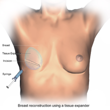
- Tissue Expander - Breast implants This is the most common technique used worldwide. The surgeon inserts a tissue expander, a temporary silastic implant, beneath a pocket under the pectoralis major muscle of the chest wall. The pectoral muscles may be released along its inferior edge to allow a larger, more supple pocket for the expander at the expense of thinner lower pole soft tissue coverage. The use of acellular human or animal dermal grafts have been described as an onlay patch to increase coverage of the implant when the pectoral muscle is released, which purports to improve both functional and aesthtic outcomes of implant-expander breast reconstruction.[5][6]
- In a process that can take weeks to months, saline solution is injected to progressively expand the overlaying tissue. Once the expander has reached an acceptable size, it may be removed and replaced with a more permanent implant. Reconstruction of the areola and nipple are usually performed in a separate operation after the skin has stretched to its final size.
- Flap reconstruction The second most common procedure uses tissue from other parts of the patient's body, such as the back, buttocks, thigh or abdomen. This procedure may be performed by leaving the donor tissue connected to the original site to retain its blood supply (the vessels are tunnelled beneath the skin surface to the new site) or it may be cut off and new blood supply may be connected.
- The latissimus dorsi muscle flap is the donor tissue available on the back. It is a large flat muscle which can be employed without significant loss of function. It can be moved into the breast defect while still attached to its blood supply under the arm pit (axilla). A latissimus flap is usually used to recruit soft-tissue coverage over an underlying implant. Enough volume can be recruited occasionally to reconstruct small breasts without an implant.


- Abdominal flaps The abdominal flap for breast reconstruction is the TRAM flap (Transverse Rectus Abdominis Myocutaneous flap) or its technically distinct variants of microvascular "perforator flaps" like the DIEP/SIEP flaps. In a TRAM procedure, a portion of the abdomen tissue group, including skin, adipose tissues, minor muscles and connective tissues, is taken from the patient's abdomen and transplanted onto the breast site. Both TRAM and DIEP/SIEP use the abdominal tissue between the umbilicus and the pubis. The DIEP flap and free-TRAM flap require advanced microsurgical technique and are less common as a result. Both can provide enough tissue to reconstruct large breasts. These procedures are preferred by some breast cancer patients because they result in an abdominoplasty (tummy tuck), and allow the breast to be reconstructed with one's own tissues instead of a foreign implant. TRAM flap procedures may weaken the abdominal wall and torso strength, but are tolerated well in most patients. To prevent muscle weakness and incisional hernias, the portion of abdominal wall exposed by reflection of the rectus abdominis muscle may be strengthened by a piece of surgical mesh placed over the defect and sutured in place. Perforator techniques such as the DIEP (deep inferior epigastric perforator) flap and SIEA (superficial inferior epigastric artery) flap require precise dissection of small perforating vessels through the rectus muscle, and purport the advantage of less weakening of the abdominal wall, though rectus abdominus muscle function may still be compromised. Other total autologous tissue breast reconstruction donor sites include the buttocks (superior or inferior gluteal artery perforator flaps (SGAP or IGAP)). The purpose of perforator flaps (DIEP, SIEA, SGAP, IGAP) is to provide sufficient skin and fat for an aesthetic reconstruction while minimizing morbidity from harvesting the underlying muscles. See free flap breast reconstruction for more information.
- Mold-assisted reconstruction Made using laser scanning and 3D printing, a patient-specific silicone mold can be used as an aid during surgery. It is used as a guide for orienting and shaping the flap to improve accuracy and symmetry.[7]
Other considerations
To restore the appearance of the breast with surgical reconstruction, there are a few options regarding the nipple and areola.
- A nipple prosthesis can be used to restore the appearance of the reconstructed breast. Impressions can be made and photographs can be used to accurately replace the nipple lost with some types of mastectomies. This can be instrumental in restoring the psychological well-being of the breast cancer survivor. The same process can be used to replicate the remaining nipple in cases of a single mastectomy. Ideally, a prosthesis is made around the time of the mastectomy and it can be used just weeks after the surgery.
- Nipple reconstruction is usually delayed until after the breast mound reconstruction is completed so that the positioning can be planned precisely. There are several methods of reconstructing the nipple-areolar complex, including:
- Nipple-Areolar Composite Graft (Sharing) - if the contralateral breast has not been reconstructed and the nipple and areolar are sufficiently large, tissue may be harvested and used to recreate the nipple-areolar complex on the reconstructed side.
- Local Tissue Flaps - a nipple may be created by raising a small flap in the target area and producing a raised mound of skin. To create an areola, a circular incision may be made around the new nipple and sutured back again. The nipple and areolar region may then be tattooed to produce a realistic colour match with the contralateral breast.
- Local Tissue Flaps With Use of AlloDerm - as above, a nipple may be created by raising a small flap in the target area and producing a raised mound of skin. AlloDerm (cadaveric dermis) can then be inserted into the core of the new nipple acting like a "strut" which may help maintain the projection of the nipple for a longer period of time. The nipple and areolar region may then be tattooed later.[8] There are however some important issues in relation to NAC Tattooing that should be considered prior to opting for tattooing, such as the choice of pigments and equipment used for the procedure.[9]
One of the challenges in breast reconstruction is to match the reconstructed breast to the mature breast on the other side (often fairly 'ptotic' - droopy.) This often requires a lift (mastopexy), reduction, or augmentation of the other breast.
Follow-up and recovery
Recovery from implant-based reconstruction is generally faster than with flap-based reconstructions, but both take at least three to six weeks of recovery and both require follow-up surgeries in order to construct a new areola and nipple. All recipients of these operations should refrain from strenuous sports, overhead lifting, and sexual activity during the recovery period. TRAM flap patients can show abdominal-muscle weakness on electromyogram (EMG) studies, but clinically most patients who have undergone unilateral breast reconstruction (reconstruction of one breast only) return to normal activities after recovery.
Patients who have undergone bilateral breast reconstruction with TRAM flaps (i.e. reconstruction of both breasts) require sacrifice of both rectus muscles and tend to have permanent abdominal strength loss. For this reason, many plastic surgeons now frown upon bilateral breast reconstruction with TRAM flaps. This also explains the significant patient interest in perforator flap techniques such as the DIEP flap which preserves abdominal muscle function long-term. These patients tend to return to full activity after several weeks without permanent limitations.
There is little information about upper body exercise post-mastectomy. Issues such as simple mastectomy, mastectomy with reconstruction, and mastectomy with lymph node excision and reconstruction all factor into limitations to amount and extent of upper body exercise. Generally, cardiac exercise (treadmill, walking, etc.) are approved for rehabilitation post-surgery and for weight control.[cita requerida]
Women who have undergone breast reconstruction must still be followed for local or regional recurrence of their cancer with manual exams of the breast/chest wall and axilla.
The most effective relief from breast reconstruction is hilotherapy, a therapy that provides relief from hematoma, pain, and swelling post-surgery without the dangers of frostbite and skin necrosis.[cita requerida]
See also
- Breast implant
- Breast lift
- Breast reduction plasty
- Free flap breast reconstruction
- Nipple prosthesis
Additional images
-
Breast Reconstruction using a Prosthesis
References
- ↑ D'Souza, N; Darmanin, G; Fedorowicz, Z (6 de julio de 2011). «Immediate versus delayed reconstruction following surgery for breast cancer.». The Cochrane database of systematic reviews (7): CD008674. PMID 21735435. doi:10.1002/14651858.CD008674.pub2.
- ↑ «Breast Reconstruction: Immediate or Delayed».
- ↑ Harcourt DM, Rumsey NJ, Ambler NR et al. (2003). «The psychological effect of mastectomy with or without breast reconstruction: a prospective, multicenter study». Plast Reconstr Surg 111 (3): 1060-8. PMID 12621175. doi:10.1097/01.PRS.0000046249.33122.76.
- ↑ Brandberg Y, Malm M, Blomqvist (2000). «SVEA," comparing effects of three methods for delayed breast reconstruction on quality of life, patient-defined problem areas of life, and cosmetic result». Plast Reconstr Surg 105 (1): 66. PMID 10626972. doi:10.1097/00006534-200001000-00011.
- ↑ Breuing KH, Warren SM (Sep 2005). «Immediate bilateral breast reconstruction with implants and inferolateral AlloDerm slings». Ann Plast Surg. 55 (3): 232-9. PMID 16106158. doi:10.1097/01.sap.0000168527.52472.3c.
- ↑ Salzberg CA (Jul 2006). «Nonexpansive immediate breast reconstruction using human acellular tissue matrix graft (AlloDerm)». Ann Plast Surg. 57 (1): 1-5. PMID 16799299. doi:10.1097/01.sap.0000214873.13102.9f.
- ↑ Ferry Melchels et al. 2011 Biofabrication 3 034114 doi 10.1088/1758-5082/3/3/034114
- ↑ Garramone, D.O., Charles E; Lam, Ben (May 2007). «Use of AlloDerm in primary nipple reconstruction to improve long-term nipple projection.». Plastic and reconstructive surgery 119 (6): 1663-8. PMID 17440338. doi:10.1097/01.prs.0000258831.38615.80.
- ↑ A. Darby. 3D Nipple Tattooing a New Service? (inc revisions). CosmeticTattoo.org Educational Articles 24/10/2013
External links
- Breast Reconstruction Following Breast Removal from the American Society of Plastic Surgeons
- National Cancer Institute breast cancer page
- US Dept. of Labor - Women's Health & Cancer Rights Act of 1998
- Plastic Surgery Stirs a Debate
Plantilla:Operations and other procedures of the integumentary system
 Wikimedia Commons alberga una categoría multimedia sobre Reconstrucción mamaria.

