
Archivo:RLS 12blauLeg.png
Tamaño de esta previsualización: 397 × 599 píxeles. Otras resoluciones: 159 × 240 píxeles · 318 × 480 píxeles · 883 × 1332 píxeles.
Ver la imagen en su resolución original (883 × 1332 píxeles; tamaño de archivo: 1,56 MB; tipo MIME: image/png)
Resumen
| Descripción |
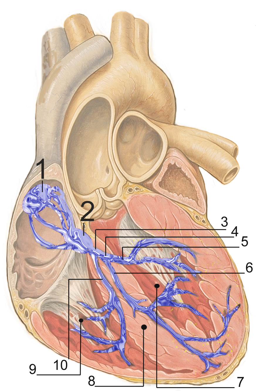
English: Electrical conduction system of the heart:
Français : Répartition du tissus nodal dans le coeur:
Русский: Проводящая система сердца:
Español: Sistema de conducción del corazón:
Deutsch: Erregungsleitungssystem:
Polski: Układ bodźcotwórczo-przewodzący:
|
| Fecha | |
| Fuente | self made, based upon Image:Heart anterior view coronal section.jpg by Patrick J. Lynch (Patrick J. Lynch; illustrator; C. Carl Jaffe; MD; cardiologist Yale University Center for Advanced Instructional Media ) |
| Autor | J. Heuser |
| Permiso (Reutilización de este archivo) |
Creative Commons Attribution 2.5 License 2007 |
preserve our creative credits: Patrick J. Lynch, medical illustrator; C. Carl Jaffe, MD, cardiologist. https://creativecommons.org/licenses/by/2.5/
Licencia
Este archivo está disponible bajo la licencia Creative Commons Reconocimiento 2.5 Genérica.
- Eres libre:
- de compartir – de copiar, distribuir y transmitir el trabajo
- de remezclar – de adaptar el trabajo
- Bajo las siguientes condiciones:
- atribución – Debes otorgar el crédito correspondiente, proporcionar un enlace a la licencia e indicar si realizaste algún cambio. Puedes hacerlo de cualquier manera razonable pero no de manera que sugiera que el licenciante te respalda a ti o al uso que hagas del trabajo.
Historial del archivo
Haz clic sobre una fecha y hora para ver el archivo tal como apareció en ese momento.
| Fecha y hora | Miniatura | Dimensiones | Usuario | Comentario | |
|---|---|---|---|---|---|
| actual | 17:51 2 mar 2007 | | 883 × 1332 (1,56 MB) | JHeuser | {{Information |Description = Heart; conduction system |Source = self made, based upon Image:Heart anterior view coronal section.jpg by Patrick J. Lynch (Patrick J. Lynch; illustrator; C. Carl Jaffe; MD; cardiologist Yale University Center for Advanc |
Usos del archivo
La siguiente página usa este archivo:
Uso global del archivo
Las wikis siguientes utilizan este archivo:
- Uso en ar.wikipedia.org
- Uso en az.wikipedia.org
- Uso en bg.wikipedia.org
- Uso en bs.wikipedia.org
- Uso en ca.wikipedia.org
- Uso en cs.wikipedia.org
- Uso en de.wikipedia.org
- Uso en el.wikipedia.org
- Uso en en.wikipedia.org
- Uso en fa.wikipedia.org
- Uso en fr.wikipedia.org
- Uso en he.wikipedia.org
- Uso en id.wikipedia.org
- Uso en ko.wikipedia.org
- Uso en lt.wikipedia.org
- Uso en lv.wikipedia.org
- Uso en ml.wikipedia.org
- Uso en or.wikipedia.org
- Uso en pl.wikipedia.org
- Uso en pt.wikipedia.org
- Uso en ru.wikipedia.org
- Uso en sh.wikipedia.org
- Uso en sk.wikipedia.org
- Uso en sr.wikipedia.org
- Uso en ta.wikipedia.org
- Uso en th.wikipedia.org
- Uso en uk.wikipedia.org
- Uso en uz.wikipedia.org
- Uso en www.wikidata.org