
Archivo:Aniksosaurus histology.png

Tamaño de esta previsualización: 440 × 600 píxeles. Otras resoluciones: 176 × 240 píxeles · 352 × 480 píxeles · 563 × 768 píxeles · 751 × 1024 píxeles · 1604 × 2187 píxeles.
Ver la imagen en su resolución original (1604 × 2187 píxeles; tamaño de archivo: 5,55 MB; tipo MIME: image/png)
Resumen
| Descripción |
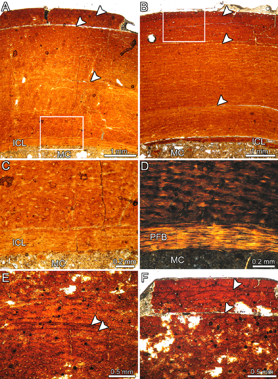
English: (slightly modified original figure caption) Bone histology of Aniksosaurus darwini (Theropoda: ?Coelurosauria) tibiae from the lower Upper Cretaceous of Patagonia. (A, B) Cyclical growth marks (arrowheads) in the cortical bone tissue of specimens MDT-PV 1/1 (A) and MDT-PV 1/28 (B). (C, D) Close-up of the inner cortex of specimen MDT-PV 1/1 (box inset in A) viewed under normal (C) and polarized (D) light. Compare the soft birefringence in some areas of the primary matrix with the strong birefringence of the ICL. (E) Double LAG (arrowheads) in the cortical tissue of specimen MDT-PV 1/1. (F) Detailed view of the outermost deposited LAGs in specimen MDT-PV 1/1. Except for (D), all figures viewed under normal light. Abbreviations: ICL, inner circumferential layer; MC, marrow cavity; PFB, parallel-fibred bone tissue. Deutsch: (modifizierte Übersetzung der Original-Bildunterschrift) Knochenhistologie fossiler Schienbeinknochen (Tibiae) von Aniksosaurus darwini (Theropoda: ?Coelurosauria) aus der unteren Oberkreide von Patagonien. (A, B) Zyklische Wachstumsmarken (Pfeile) im kortikalen Knochengewebe von Exemplar MDT-PV 1/1 (A) und MDT-PV 1/28 (B). (C, D) Vergrößerung der inneren Kortikalis von Exemplar MDT-PV 1/1 (der in A eingekästelte Bereich) unter normalem (C) und polarisiertem (D) Licht. Man beachte die geringe Doppelbrechung in einigen Bereichen der primären Matrix im Vergleich zur hohen Doppelbrechung der inneren Randschicht (engl.: inner circumferential layer, hier abgek. ICL). (E) Doppelte Wachstumslamelle (engl.: line of arrested growth, allg. abgek. LAG, wörtlich: ‚Linie gestoppten/gebremsten Wachstums‘; siehe Pfeile) im kortikalen Knochengewebe von Exemplar MDT-PV 1/1. (F) Detailansicht der am weitesten außen liegenden Wachstumslamellen bei Exemplar MDT-PV 1/1. Mit Ausnahme von (D), sind alle Bilder unter normalem Licht aufgenommen. Abkürzungen: ICL, innere Randschicht der Kortikalis; MC, Markhöhle; PFB, parallelfaseriges Knochengewebe. |
| Fecha | |
| Fuente | fig. 9 in: The Behavioral Implications of a Multi-Individual Bonebed of a Small Theropod Dinosaur. PLoS ONE 8(5): e64253. doi:10.1371/journal.pone.0064253 |
| Autor | Lucio M. Ibiricu, Rubén D. Martínez, Gabriel A. Casal, Ignacio A. Cerda |
Licencia
|
Este archivo está disponible bajo la licencia Creative Commons Reconocimiento 2.5 Genérica.
|
This file was published in a Public Library of Science journal. Their website states that the content of all PLOS journals is published under the Creative Commons Attribution 4.0 license (or its previous version depending on the publication date), unless indicated otherwise.
|
Historial del archivo
Haz clic sobre una fecha y hora para ver el archivo tal como apareció en ese momento.
| Fecha y hora | Miniatura | Dimensiones | Usuario | Comentario | |
|---|---|---|---|---|---|
| actual | 01:12 28 may 2013 | | 1604 × 2187 (5,55 MB) | Ras67 | cropped |
| 12:34 17 may 2013 |  | 1621 × 2204 (7,41 MB) | FunkMonk | {{Information |Description=Bone histology of Aniksosaurus darwini tibiae. (A, B) Cyclical growth marks (arrowheads) in the cortical bone tissue of specimens MDT-PV 1/1 (A) and MDT-PV 1/28 (B). (C, D) Close-up of the inner cortex of specimen MDT-PV 1/1... |
Usos del archivo
La siguiente página usa este archivo:
Uso global del archivo
Las wikis siguientes utilizan este archivo:
- Uso en de.wikipedia.org
- Uso en en.wikipedia.org