
Archivo:Nanotubes.png

Tamaño de esta previsualización: 782 × 600 píxeles. Otras resoluciones: 313 × 240 píxeles · 626 × 480 píxeles · 1002 × 768 píxeles · 1280 × 982 píxeles · 2128 × 1632 píxeles.
Ver la imagen en su resolución original (2128 × 1632 píxeles; tamaño de archivo: 2,29 MB; tipo MIME: image/png)
Resumen
| Descripción |
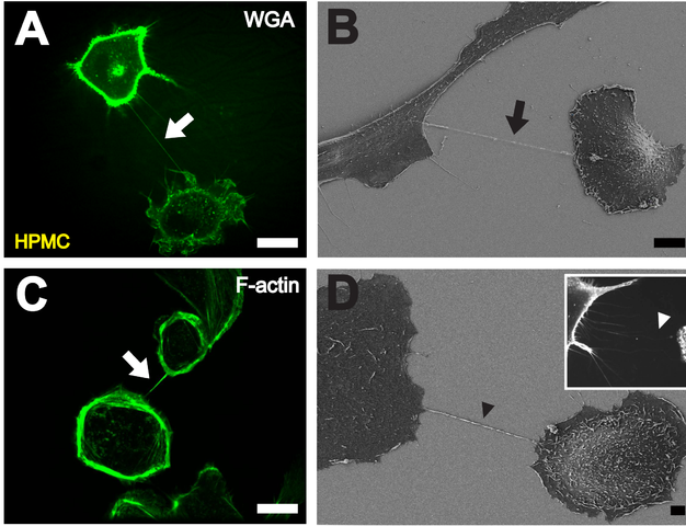
English: A. High resolution 3D live-cell fluorescence image of a NT (white arrow) connecting two primary mesothelial cells one hour after plating on a collagen I coated glass cover slide. To facilitate detection, cell membranes were stained with WGA Alexa Fluor® 488. Scale bar: 20 µm. B Depiction of a NT (black arrow) between two cells with scanning electron microscopy one hour after cell plating. Scale bar: 10 µm. C F-actin staining by fluorescently labeled phalloidin showing actin being present in NTs between individual HPMCs (white arrow). Scale bar: 20 µm. D Scanning electron microscope picture of a substrate-associated filopodia-like extension as potential NT precursor (black arrowhead). The insert shows a fluorescence microscopic image of substrate associated filopodia-like protrusions approaching a neighboring cell (white arrowhead). Scale bar: 2 µm. |
| Fecha | |
| Fuente | PLoS One |
| Autor | Ranzinger J, Rustom A, Abel M, Leyh J, Kihm L, et al. |
Licencia
|
Este archivo está disponible bajo la licencia Creative Commons Reconocimiento 2.5 Genérica.
|
This file was published in a Public Library of Science journal. Their website states that the content of all PLOS journals is published under the Creative Commons Attribution 4.0 license (or its previous version depending on the publication date), unless indicated otherwise.
|
Historial del archivo
Haz clic sobre una fecha y hora para ver el archivo tal como apareció en ese momento.
| Fecha y hora | Miniatura | Dimensiones | Usuario | Comentario | |
|---|---|---|---|---|---|
| actual | 11:44 28 dic 2011 | | 2128 × 1632 (2,29 MB) | Gustavocarra | {{Information |Description ={{en|1='''A'''. High resolution 3D live-cell fluorescence image of a NT (white arrow) connecting two primary mesothelial cells one hour after plating on a collagen I coated glass cover slide. To facilitate detection, cell me |
Usos del archivo
La siguiente página usa este archivo:
Uso global del archivo
Las wikis siguientes utilizan este archivo:
- Uso en ar.wikipedia.org
- Uso en en.wikipedia.org
- Uso en gl.wikipedia.org
- Uso en ja.wikipedia.org
- Uso en pt.wikipedia.org